


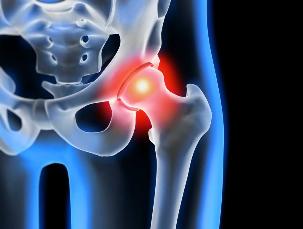
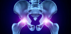
Osteoarthritis of the hip — degenerative-degenerative disease, that is characterized by destruction of hyaline cartilage. The disease develops gradually, accompanied by a syndrome of pain and reduction of volume of movements. In the absence of intervention in the initial phase of arthritis, a few years later occurs the atrophy muscle femoral. Damaged, the limb is shortened, and the remodeling of the joint space leads to partial or complete paralysis of the hip. The causes of disease become injury pre-existing curvature of the spine, systemic diseases of the musculoskeletal system.
Osteoarthritis is usually diagnosed in patients of middle age and the elderly. The diagnosis will be issued on the basis of the results of instrumental examinations — x-ray, mri, CT, arthroscopy. The treatment of the disease of 1 and 2 degree of severity, conservative. When it is detected ankylosis or ineffectiveness of medical therapy carries out surgical intervention (arthrodesis, prosthesis).
The mechanism of development of pathology
The hip joint form two bones — the iliac and the femur. The lower section of the iliac bone presented his body, which is involved in a plexus with the femur is the bone that forms the superior part of the acetabulum. During the movement of the articular fossa of the property, and the femoral head moves freely. What is "articulate" device, the hip joint allows you to bend, stretch, turn, promotes abstraction, bring the thighs. Smooth sliding of the articulated structures provides smooth, elastic, elastic, hyaline cartilage, along the acetabulum and the femoral head. Its main functions — redistribution of the loads in movement, alert, quick wear and tear of bone tissues.
Under the influence of internal or external factors, broken trophic cartilage. Does not have a circulatory system — nutrients, fabric provides the synovial fluid. With osteoarthritis thickens, it becomes viscous. The nutrient deficiency causes drying of the surface of hyaline cartilage. She is covered with cracks, which results in a constant micro lesions of tissues in flexion or extension of the hip. The cartilage thinning, lose their depreciation of property. To "adapt" to an increase of pressure, the bones are deformed. And in the background of the deterioration of the metabolism in the tissues are advancing the destructive-degenerative changes.
Causes and triggers
Idiopathic or primary osteoarthritis develops without any reason. It is believed that the destruction of the cartilage tissue occurs due to the natural aging of the body, slow down the reconstruction of processes, reduction of the production of collagen and other compounds necessary for a complete regeneration of the structures of the hip joint. Secondary osteoarthritis occurs on a background already present in the body of the pathological state. The most common reason secondary of the disease are:
- previous injury — damage to the sheath of the tendons of the machine, tearing the muscle, their total detachment from the bone marrow of the base, fractures, dislocations;
- the violation of the development of the joint, the defects of dysplastic disorders;
- autoimmune disease — rheumatoid, reactive, psoriatic arthritis, systemic lupus erythematosus;
- nonspecific inflammatory diseases, such as arthritis purulent;
- the specific infections — gonorrhea, syphilis, brucellosis, ureaplasmosis, trichomoniasis, tuberculosis, osteomyelitis, encephalitis;
- violation of the functioning of the endocrine system;
- degenerative-dystrophic pathology — osteochondropathy head of the femur;
- hypermobility of the joints, due to the production of "excess supply" of collagen, causing an excess of them, of mobility, weakness of the ligaments.
As well as the reason for the development of osteoarthritis can become hemarthrosis (bleeding into the cavity of the hip), the causing factors relate to violations of the blood. The conditions at the onset of the disease are excess weight, excessive exercise, sedentary lifestyle. Its development and lead the organization to the wrong athletes during the workouts, the lack in the diet of foods with a high content of trace elements, giraud and water-soluble vitamins. Post-operative osteoarthritis occurs after a couple of years after the surgical intervention, especially if it is accompanied by excising large amounts of tissue. Osteoarthritis of the hip can not be inherited. But the presence of some defects of characteristics (violation of metabolism, the structure of the skeleton), the probability of development is significantly increased.
The symptoms
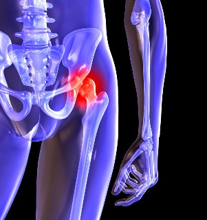
The main symptoms of osteoarthritis of the hip pain when walking in an area of the hip, knee, joint. A man who is suffering from stiffness of movement, stiffness, especially in the early hours of the morning. To stabilize the joint, the patient begins the lame from, change its gait. With the passage of time due to muscle atrophy and deformation of the joint, the limb remarkably shortened. Another feature of pathology — limitation of the seizure of the hip. For example, the difficulties arise when trying to sit down on a stool, legs splayed to the sides.
Also "launched" OSTEOARTHRITIS can be cured at home! Just don't forget of times per day to spread this...
For osteoarthritis of first degree of severity, characterized by recurring pain that occur after intense physical efforts. They are located in the area of articulation and disappear after a long period of rest.
With osteoarthritis of second degree of hip link the manifestation of the pain increases. The sensations of discomfort occur even in the resting state, shall apply to the thigh and groin, are amplified during the lifting of weights, or an increase in locomotor activity. To eliminate the hip pain, the man begins to barely limp. Note the limitation of joint movements, in particular allocation mechanisms and internal rotation of the hip.
Osteoarthritis of the third degree is characterized by continuous severe pain. When you ride you experience any problems, then when you walk a man forced to use a cane or crutches. Because of the weakness of the exhaust the muscle femoral occurs the displacement of the bones in the pelvic in the frontal plane. For the payment of accident shortening of the diseased leg when traveling, you lean toward the limb the patient. This causes a strong shift of the gravity centre and the increased load of the joint. At this stage of the arthrosis develops expressed ankylosis of the joint.
| Measure | Radiographic signs |
| The first | The changes are not expressed in a dramatic way. The articular slit moderately irregular restricted, there is no destruction of the surface of the femur. Internal or external edge of the acetabulum, there are small growths of bone |
| The second | The height of the joint space greatly reduced due to its irregular remodeling. Bone the head of the femur is biased upward, deformed, is increased, the contours become jagged. Osteophytes are formed on the inner and outer surface of the edges of the articular fossa |
| The third | We see, in total or in part, the remodeling of the joint space. The head of the femur is very extended. Multiple osteophytes are found on all surfaces of the acetabulum |
Diagnostics
When you make the diagnosis, the physician takes into account the clinical manifestations of the disease, the history, the results of the external examination of the patient and research tools. The most informative x-ray. With its help, you assessed the state of the hip, is set to stream stage the degree of lesion of the cartilage tissues, and in some cases the reason for the development. If the cervical-diaphyseal most common is increased, and the acetabulum is smoothed and flattened, with large probability one can assume dysplastic defects changes of the joint. For the perthes disease: indicates broken bone shape of the hip. The x-ray allows you to vyvit post-traumatic osteoarthritis, despite the lack of a previous history of disease injury. Are also used other diagnostic methods:
- The TC allows you to detect the proliferation of the edges of the bony plates, the osteophytes;
- Magnetic RESONANCE imaging performed for the evaluation of the state of the connective tissue of the structures and their degree of involvement in the pathological process.
If necessary, the inner surface of the joint surveys with the help of arthroscopic instruments. The differential diagnosis is carried out the elimination of knee osteoarthritis, lumbosacral, or osteoarthritis. The pain from osteoarthritis can masquerade as the clinical manifestations of create a syndrome caused by the violation or the inflammation of the nerve. To exclude neurogenic pathology usually succeeds with the help of a series of tests. Osteoarthritis of the hip is necessarily different from the top of bursitis of the hip, ankylosing Spondylitis, reactive arthritis. To exclude autoimmune diseases are conducted biochemical studies of blood and synovial fluid.
Surgery
The ineffectiveness of conservative therapy and the complicated diagnosis pathology takes place. To restore the cartilage tissue in the knee, damaged in osteoarthritis, without the intervention of the prosthesis impossible, but with the right approach to treatment, compliance with all medical requirements, managed by a healthy lifestyle, exercise classes, medical, regular massage courses, taking vitamins and proper diet you can stop the process of destruction, and the destruction of the cartilage and the joints of the hip.